Vladislav, 18, and the effort to save his natural tooth

At 18, Vladislav already had several teeth with extensive decay. In one tooth, the decay had reached the pulp and destroyed so much of the tooth that a dentist in Switzerland recommended extraction. His mother worried about him losing a permanent tooth at such a young age and wanted to find out whether it could be saved.
On the recommendation of friends, Vladislav and his mother travelled from Switzerland to Lilladent in Budapest. She chose the clinic because it focuses on preserving natural teeth with modern restorative techniques and had Russian-speaking staff who could help them communicate during the consultation.
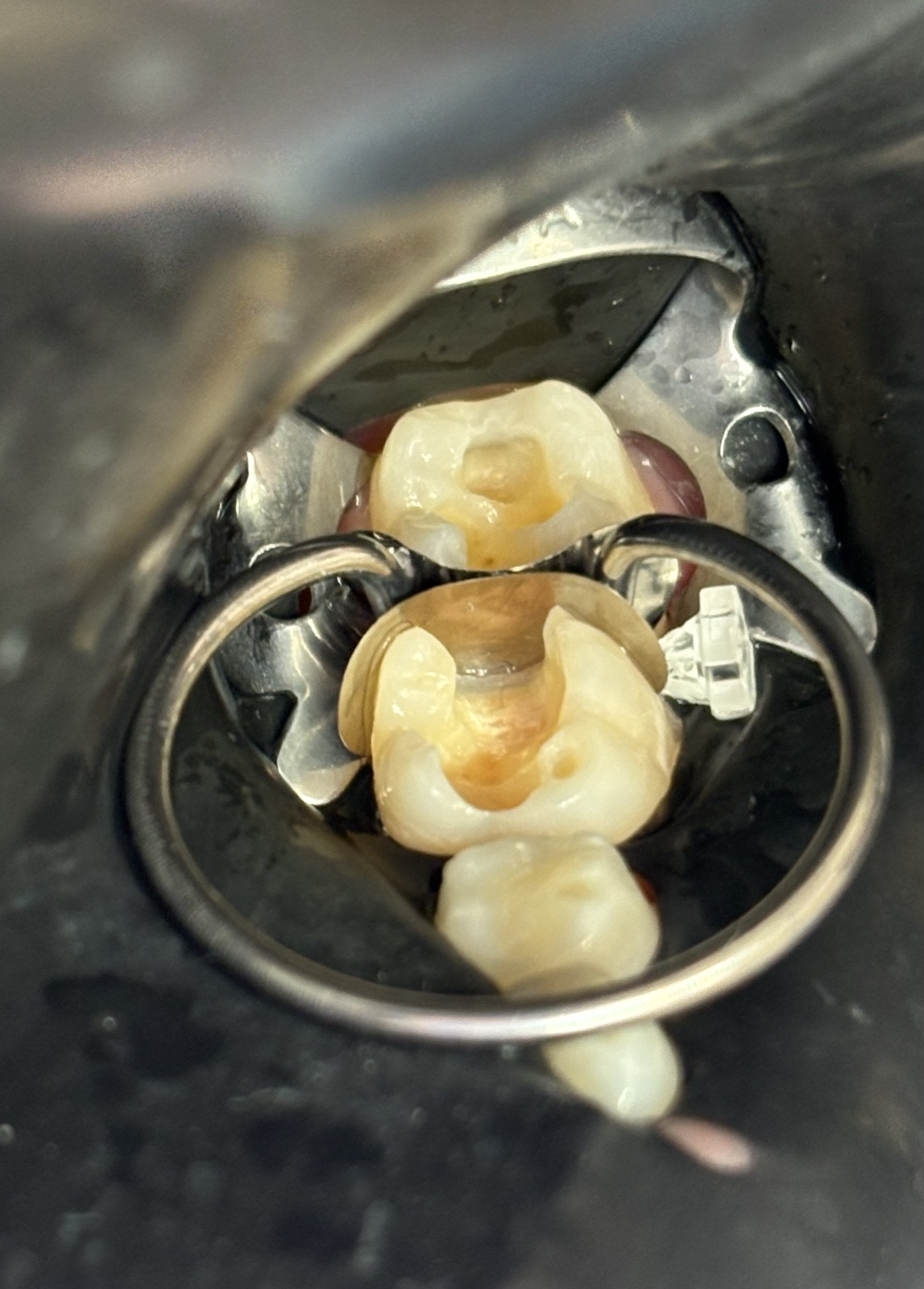
At the examination, we found that the tooth was severely damaged, but enough healthy structure remained to restore it. One challenge was that the decay extended well below the gumline. With Deep Margin Elevation, we used composite to raise the edge of the restoration to a more accessible position. The plan was to raise that margin, then carry out root canal treatment and fit a ceramic crown to protect the remaining tooth structure.
Vladislav also had several other cavities that needed treatment and two front teeth that tilted inward, affecting the appearance of his smile. After we explained the options, he and his mother decided to proceed at Lilladent. They could have the natural tooth saved using modern restorative techniques, and the cost suited their family.
The following photos show the stages of Vladislav’s treatment at Lilladent.
Photos before and after treatment
1. Incisors before and after restoration


The first photo shows the incisors before treatment. The next two show Vladislav’s two incisors after restoration with direct composite veneers.
Saving tooth 46
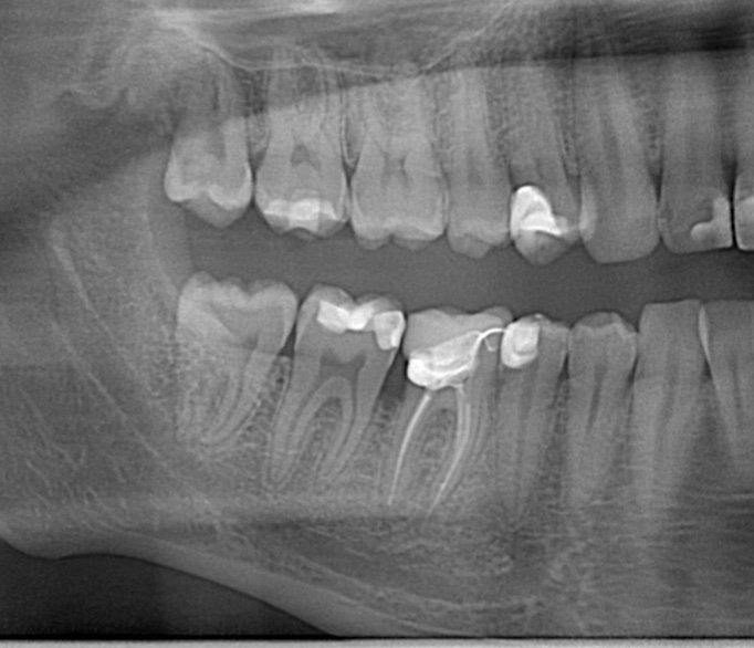
2. Tooth 46 before, during, and after treatment
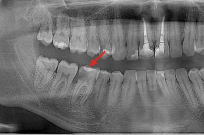
Before treatment. Tooth 46 (red arrow) had a large cavity and inflammation around the alveolar bone crest. Extraction had previously been recommended.
After treatment. The tooth was treated with root canal therapy, deep margin elevation, and an e.max ceramic crown. The inflammation around the alveolar bone crest resolved.
Clinical photos during and after treatment


The first photo shows tooth 46 during treatment. The second photo shows it after the e.max crown was fitted.
Restoring the molars
3. Restoration of teeth 36 and 37